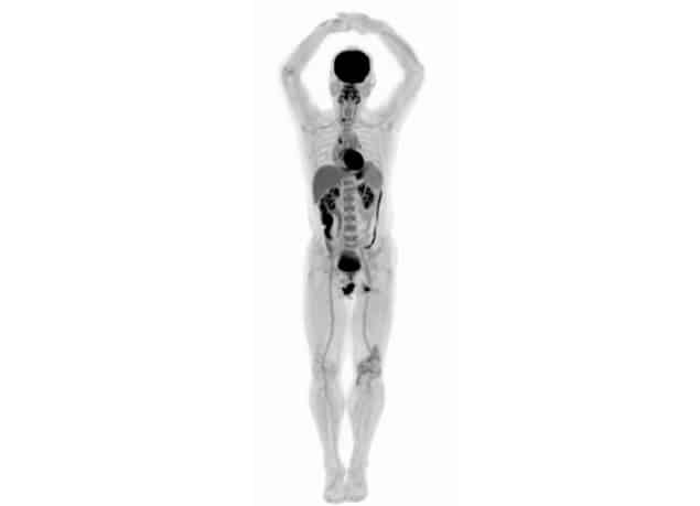
EXPLORER PET/CT produces first total-body scans

The brainchild of UC Davis scientists Simon Cherry and Ramsey Badawi, EXPLORER has a far higher sensitivity than current commercial scanners and can produce a whole-body diagnostic scan in as little as 20-30 s. It can also create movies that track radiolabelled drugs as they move around the body. The machine can scan up to 40 times faster, or use up to 40 times less radiation dose, than current PET scans, making it possible to conduct repeated studies in an individual, or dramatically reduce dose in paediatric studies.
“The trade-off between image quality, acquisition time and injected radiation dose will vary for different applications, but in all cases, we can scan better, faster or with less radiation dose, or some combination of these,” Cherry explains.
Imaging first
Badawi and Cherry first conceptualized the total-body scanner 13 years ago. In 2011, a $1.5 million grant from the National Cancer Institute allowed them to establish a consortium of researchers and other collaborators. And in 2015, a $15.5 million grant from the NIH enabled them to team up with a commercial partner and build the first EXPLORER scanner.The first human images were acquired in collaboration with Shanghai-based United Imaging Healthcare (UIH) — which built the system and will manufacture the scanner — and Zhongshan Hospital.
“While I had imagined what the images would look like for years, nothing prepared me for the incredible detail we could see on that first scan,” says Cherry. “While there is still a lot of careful analysis to do, I think we already know that EXPLORER is delivering roughly what we had promised.”
The team used the scanner to image the delivery and distribution of fluorodeoxyglucose in real time. The resulting movie shows how, in the first few seconds after injection into a leg vein, the tracer travelled to the heart and was then distributed through the arteries to all organs in the body. Gradual accumulation of the glucose could be seen in the heart, brain and liver.
Read more

Developing the world’s first full-body PET scanner


 https://youtu.be/thGvKuqDPDE

No comments:
Post a Comment
Thank you for your feedback.